Article Plan⁚ X-Ray Positioning Chart with Images PDF
This article explores X-ray positioning charts featuring images in PDF format. It delves into forearm, elbow, and humerus radiographic projections, incorporating precise patient positioning. Skull positioning and range of motion measurements are examined, emphasizing diagnostic radiology and efficiency. We’ll cover creating a comprehensive chart with guide wire placement.
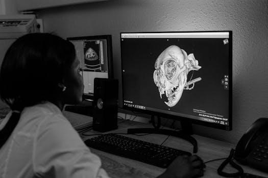
X-ray positioning charts are indispensable tools in the field of diagnostic radiology, serving as visual guides for radiographers and technicians. These charts provide detailed instructions and illustrations on how to accurately position patients for various radiographic examinations, ensuring optimal image quality and diagnostic accuracy. The use of X-ray positioning charts is crucial for obtaining clear and informative images that aid in the diagnosis and treatment of medical conditions.
These charts typically include diagrams, photographs, or illustrations that demonstrate the correct patient positioning, beam alignment, and exposure parameters for specific anatomical regions. They often cover a wide range of projections, including anterior-posterior (AP), posterior-anterior (PA), lateral, and oblique views, as well as specialized projections for specific clinical indications.
The primary goal of X-ray positioning charts is to standardize radiographic procedures, reduce errors, and improve the consistency of image acquisition. By following the guidelines outlined in these charts, radiographers can minimize patient exposure to radiation, reduce the need for repeat examinations, and ensure that the images obtained are of diagnostic quality. In essence, X-ray positioning charts are pivotal in enhancing the efficiency and effectiveness of radiographic imaging practices in healthcare settings.
Importance of Accurate Patient Positioning in Radiography
Accurate patient positioning is paramount in radiography for several critical reasons. Firstly, precise positioning ensures that the anatomical region of interest is clearly visualized, allowing for accurate diagnosis. Incorrect positioning can lead to distortion, superimposition of structures, or incomplete visualization, potentially obscuring pathology or mimicking other conditions. This underscores the need for meticulous attention to detail during patient preparation and positioning.
Secondly, accurate positioning minimizes patient exposure to radiation. When the patient is correctly positioned, fewer repeat examinations are required, reducing the cumulative radiation dose. This is especially important for vulnerable populations such as children and pregnant women. By adhering to standardized positioning techniques, radiographers can optimize image quality while minimizing radiation risk.
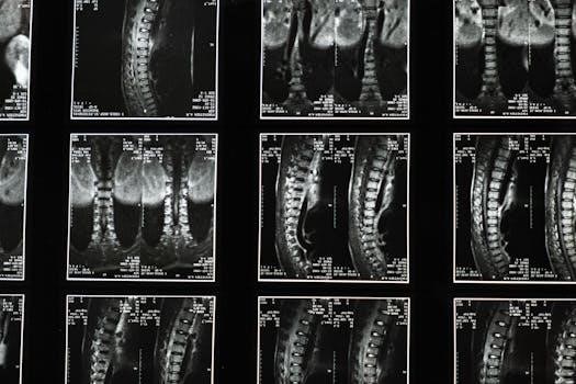
Thirdly, consistent and accurate positioning contributes to the reproducibility of radiographic examinations. This is essential for monitoring disease progression, evaluating treatment response, and comparing images over time. Standardized positioning allows radiologists to confidently assess changes in anatomical structures and identify subtle abnormalities. In conclusion, accurate patient positioning is fundamental to diagnostic accuracy, radiation safety, and the overall quality of radiographic imaging.
Common X-Ray Projections and Positioning Techniques

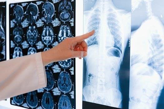
Radiography employs various projections and positioning techniques to visualize different anatomical structures effectively. Anteroposterior (AP) and Posteroanterior (PA) projections are frequently used for chest and abdominal imaging. AP views, as seen in stress X-rays, involve positioning the patient with the anterior aspect facing the X-ray tube, while PA views have the posterior aspect facing the tube. Lateral projections, where the X-ray beam passes through the patient from side to side, are essential for evaluating skeletal structures and soft tissues.
Oblique projections are used to visualize specific structures by angling the patient relative to the X-ray beam. These are particularly useful for examining joints and detecting subtle fractures. Axial projections, such as the axial view of the foot, provide a cross-sectional perspective of the anatomy.
Positioning techniques vary depending on the projection and anatomical region. Proper collimation, centering of the X-ray beam, and immobilization are crucial for minimizing radiation exposure and ensuring image quality. Utilizing positioning aids and clear communication with the patient are also essential for achieving accurate and reproducible results. Guide templates can aid in precise positioning, especially in surgical planning.
Forearm, Elbow, and Humerus Radiographic Projections
Radiographic evaluation of the forearm, elbow, and humerus involves specific projections to visualize these bony structures and surrounding soft tissues. The standard projections typically include Anteroposterior (AP) and lateral views. For the forearm, the AP view visualizes the radius and ulna, ensuring the entire forearm is included, from wrist to elbow. The lateral view is obtained with the forearm in a true lateral position, with the elbow flexed at 90 degrees.
Elbow projections include AP, lateral, and oblique views. The AP view demonstrates the distal humerus, proximal radius, and ulna. The lateral view, similar to the forearm, requires a 90-degree elbow flexion. Oblique views can help visualize specific fractures or joint abnormalities.
Humerus imaging involves AP and lateral projections, ensuring the shoulder and elbow joints are included. The AP view displays the entire humerus, while the lateral view requires rotating the arm internally or externally to obtain a true lateral projection. Proper patient positioning and collimation are essential for optimal image quality and minimizing radiation exposure. These projections, detailed in X-ray positioning charts with images in PDF format, are vital for diagnosing fractures, dislocations, and other musculoskeletal conditions.
Skull Positioning and Range of Motion Measurements

Skull radiography necessitates precise positioning to accurately visualize cranial structures. Common projections include the AP, lateral, and Towne’s views. The AP view, often with variations like the Caldwell or Waters projection, demonstrates specific facial bones and sinuses. The lateral view provides a comprehensive overview of the skull, including the sella turcica and cranial vault. Towne’s view visualizes the occipital bone and foramen magnum.
Accurate positioning ensures minimal distortion and optimal visualization of anatomical landmarks. Range of motion measurements, while less common in standard skull radiography, can be relevant in assessing craniocervical junction stability or temporomandibular joint (TMJ) function. These measurements typically involve assessing flexion, extension, lateral bending, and rotation.
X-ray positioning charts with images in PDF format are invaluable for guiding radiographers in achieving correct positioning. The charts detail specific angles, centering points, and patient instructions for each projection. Proper collimation and shielding are crucial to minimize radiation exposure. While advanced imaging modalities like CT and MRI have largely replaced skull radiography for detailed assessments, understanding basic skull projections and positioning remains fundamental in radiology practice and for initial trauma evaluations. The positioning charts aid in consistent and accurate image acquisition.
Creating a Comprehensive X-Ray Positioning Chart
Developing a comprehensive X-ray positioning chart requires meticulous attention to detail and a thorough understanding of radiographic principles. The chart should encompass a wide range of projections, catering to various anatomical regions and clinical indications. Each projection entry must include clear, concise instructions on patient positioning, part positioning, and central ray angulation. Illustrations, ideally photographs or diagrams, are essential for visual clarity and to minimize ambiguity.
The chart should specify the appropriate collimation settings to reduce scatter radiation and improve image quality. Exposure factors, such as kVp and mAs, should be provided as a starting point, recognizing that adjustments may be necessary based on patient size and tissue density. Inclusion of anatomical landmarks assists radiographers in accurately centering the X-ray beam.
A well-designed chart should also address common positioning errors and offer troubleshooting tips. Information on proper shielding techniques to minimize radiation exposure to patients and personnel is paramount. The chart’s layout should be user-friendly, with a logical organization and easy-to-read font. Consider incorporating a color-coding system to differentiate between projections or anatomical regions. Finally, the chart should be regularly reviewed and updated to reflect current best practices and technological advancements. A PDF format ensures easy distribution and accessibility.
Role of Images in X-Ray Positioning Charts
Images play a pivotal role in X-ray positioning charts, significantly enhancing their clarity, usability, and effectiveness. A high-quality image provides visual confirmation of proper patient and part positioning, which is crucial for accurate radiographic examinations. These images serve as a visual guide, demonstrating the correct anatomical alignment and beam centering required for each projection.
Including radiographic images of the final X-ray result alongside the positioning instructions offers a valuable reference point. This allows radiographers to compare their images with the ideal outcome, identifying potential errors in positioning or exposure factors. The images should clearly depict relevant anatomical landmarks, aiding in precise beam alignment and minimizing the need for repeat exposures.
Furthermore, images can illustrate common positioning mistakes, highlighting areas where errors frequently occur. By visually demonstrating these pitfalls, radiographers can proactively avoid them, improving image quality and reducing patient radiation dose. Detailed images also assist in training new radiographers, providing a practical and easily understandable learning tool. The use of images, especially in a readily accessible PDF format, makes X-ray positioning charts an indispensable resource in any radiology department.
Benefits of Using PDF Format for X-Ray Positioning Charts
The PDF format offers numerous advantages for X-ray positioning charts, making it an ideal choice for distribution and accessibility. One key benefit is its platform independence, ensuring that the chart can be viewed consistently across various operating systems and devices, be it a computer, tablet, or smartphone. This eliminates compatibility issues and allows radiographers to access the information regardless of their location or equipment.
PDFs are also easily printable, maintaining formatting and image quality when hard copies are needed. This is crucial in clinical settings where quick reference guides are often required at the point of care. The format supports embedded images and vector graphics, ensuring that the visual aids are clear and detailed, essential for precise positioning guidance. Furthermore, PDFs can be password-protected, safeguarding sensitive information and ensuring that only authorized personnel can access the charts.
The relatively small file size of PDFs makes them easy to share via email or store on network drives, facilitating efficient distribution and collaboration among radiology staff. PDF format also allows for easy indexing and searching of content, making it quick to locate specific positioning techniques or anatomical landmarks. This efficient accessibility ultimately contributes to improved workflow and reduced examination times.
Resources for X-Ray Positioning Charts with Images PDF
Radiology professionals seeking comprehensive X-ray positioning charts in PDF format with illustrative images have access to a variety of valuable resources. Many hospital radiology departments maintain internal libraries of standardized protocols, often available as PDFs, tailored to their specific equipment and patient demographics. These internal resources may include detailed positioning guides for common and specialized radiographic examinations, ensuring consistency and quality within the department.
Several professional organizations, such as the American Society of Radiologic Technologists (ASRT) and the Radiological Society of North America (RSNA), offer educational materials, including positioning guides, some of which may be available for download in PDF format. Academic institutions with radiography programs often provide online resources for students and practicing technologists. These resources may consist of lecture notes, instructional videos, and downloadable positioning charts.
Online medical imaging databases and repositories may also contain relevant PDF documents; These resources are particularly helpful for accessing less common or specialized positioning techniques; Always verify the accuracy and reliability of information obtained from online sources by cross-referencing with established textbooks and professional guidelines. Remember to consider copyright and usage restrictions when accessing and utilizing these resources.